Cornea, Corneal Therapeutics
New Therapeutic Pathways in Keratoconus
Future combination treatments could improve outcomes and reduce risks.

Ongoing research and technological advances in biomarking and genetic therapies are paving the way for innovative treatments that could transform keratoconus in the near future, according to Koji Kitazawa MD, PhD.
“Significant challenges remain, but we are making steady progress. Biomarkers play a pivotal role in the diagnosis, monitoring, and potential treatment of keratoconus—while gene therapy holds promise as a potential cure for keratoconus by addressing the genetic causes of the disease,” Dr Kitazawa said.
Dr Kitazawa defined biomarkers as measurable indicators of a biological condition or state.
“In the context of disease, they can be substances or processes indicating the presence, stage, or progression of a disease,” he explained. “Biomarkers can identify keratoconus early, even before significant symptoms appear. They can help with monitoring disease progression and the effectiveness of the treatment. Biomarkers also allow for a more personalised medical approach, leading to better clinical outcomes and reduced side effects.”
These biomarkers can be broken down into three broad subcategories: structural, proteomic, and genetic.
“Structural biomarkers identify changes detected through imaging techniques such as corneal topography or tomography that show characteristic thinning or bulging of the cornea,” he said. “Proteomic biomarkers show protein expression levels that differ between healthy and keratoconus-affected corneas, and genetic biomarkers show variations or mutations in specific genes associated with keratoconus.”
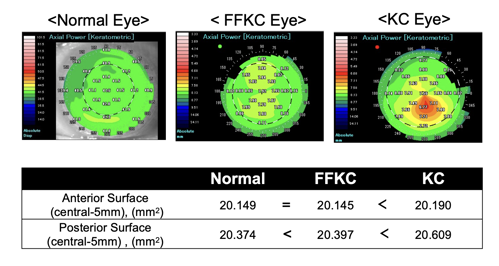
To highlight the potential utility of structural biomarkers in keratoconus, Dr Kitazawa and team evaluated the diagnostic performance of the different indices obtained between normal eyes and keratoconic eyes using anterior segment optical coherence tomography.
“Our findings revealed the anterior surface and posterior surface ratio was smaller in forme fruste keratoconus and keratoconic eyes, suggesting—theoretically—that the posterior surface area became enlarged compared to the anterior surface at the initial stage of the disease,” he said.
Proteomic biomarkers can be obtained using tear film analysis, which is minimally invasive and allows clinicians to monitor upregulation or downregulation of particular proteins for possible disease progression.
However, it is in the domain of genetic biomarkers that research progress has perhaps been the most striking in recent years, said Dr Kitazawa.
“Some reports have identified multiple loci associated with central corneal thickness and keratoconus in European and Asian populations,” he said. “Identification of these gene mutations brings into focus the potential for gene therapy for keratoconus for diagnosis and progression and paving the way to eventual gene therapy, whether in the form of gene editing, gene replacement, or gene silencing techniques.”
Multiple challenges remain in the optimal delivery method and safety of any potential gene therapy treatment, but Dr Kitazawa believes there are good grounds for optimism with each research breakthrough.
“In an ideal future scenario, the patient would go to the hospital for genetic analysis and biomarkers, and then we would integrate gene therapy with biomarker-guided therapies. We also envisage combination therapies incorporating gene therapy with other treatments such as corneal cross-linking or biomarker-guided therapies for a comprehensive approach,” he observed. “The ideal goal would be to offer more personalised medicine tailoring gene therapy to individual genetic profiles, enhancing efficiency and reducing risks.”
Dr Kitazawa presented at EuCornea 2024 in Paris.
Koji Kitazawa MD, PhD is Assistant Professor at Kyoto Prefectural University of Medicine, Kyoto, Japan. kkitazaw@koto.kpu-m.ac.jp